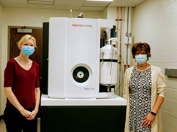
Clara Kielkopf, Ph.D., left, and Laura Calvi, M.D., stand by the University's new cryo-microscope
With the recent acquisition of Nobel Prize-winning technology and two new grants, Wilmot Cancer Institute researchers are streamlining their investigations into a malignant blood disease known as MDS, working toward discovering targeted therapies.
Laura Calvi, M.D., and Clara Kielkopf, Ph.D., are leading collaborative teams that will be using a device at the University of Rochester Medical Center — a cryo-electron microscope — that has ushered in a new era in biochemistry. The microscope allows scientists to see 3D snapshots and more details of living molecules than ever before, down to near-atomic resolution, to understand disease and uncover new ways to design drugs. The developers of the technology were awarded the Nobel Prize in Chemistry 2017.
Calvi and Kielkopf each received Edward P. Evans Foundation awards totaling $1.2 million for this project. Evans grants go to scientists seeking a cure for myelodysplastic syndromes (MDS), which originates in the bone marrow and disrupts healthy blood cell formation. MDS often leads to leukemia.
The Kielkopf lab will use the cryo-electron microscope to obtain 3D views of recurrent MDS mutations as guides for targeting molecular therapies. The modern microscope is the first of its kind in the Rochester region, Kielkopf said, and will be accessible to all UR researchers through the Electron Microscopy Shared Resource Laboratory. She is a professor of Biochemistry and Biophysics in the Center for RNA Biology.