UR Medicine / Imaging / Specialties / Cardiothoracic Imaging
Cardiothoracic Imaging

UR Medicine Cardiothoracic Imaging provides comprehensive noninvasive evaluation of both the pulmonary and cardiovascular systems. Our physicians are dedicated to the safe performance of clinical imaging exams and interpreting these exams with a subspecialty level of experience and knowledge. Our consultation is critical in the clinical evaluation of the diseases and disorders of the heart and lungs.
Thoracic imaging offers advanced imaging techniques for the evaluation of chest diseases in addition to routine assessment of the lungs with chest x-ray and chest CT. These exams include:
- CT oncologic imaging
- Lung cancer screening
- 4D CT evaluation of parathyroid adenoma
- CT/MR evaluation of thymoma
- HRCT evaluation of interstitial lung disease
- Pre-LVAD evaluation
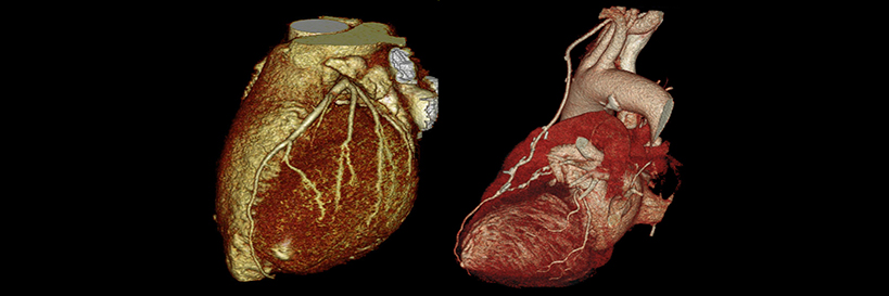
The cardiovascular subspecialists utilize the latest magnetic resonance and computed tomography technology for the noninvasive imaging of the heart and vascular system in adults and, in some cases, children.
Examples of advanced imaging vascular consultation include:
- CT and MR angiography of the thoracic aorta
- CT and MR angiography of the pulmonary arterial tree
- CT and MR Pulmonary venous anatomy evaluation (in patients with atrial fibrillation)
- CT evaluation during TAVI workup
- CT evaluation of SVC syndrome
Examples of advanced imaging cardiology consultation include:
- MR and CT evaluation of congenital heart disease
- MR evaluation of adult acquired heart disease
- CT coronary artery evaluation
Clinical areas of particular interest to the faculty include resident education, improved utilization of thoracic MRI, the incorporation of MRI in pre-TAVI evaluation, and CT quality improvement.
Our Faculty

Sean Cleary, M.D.
Assistant Professor

Joseph (Jay) Mazikas, P.A.-C
Physician Assistant