Abdominal Imaging

Using the latest technology, UR Medicine Abdominal Imaging is one of the most involved specialty services in the Department of Imaging. We concentrate on every aspect of cross-sectional imaging including CT, MRI, and ultrasonography.
Please review our Abdominal Imaging patient brochures for more details about the services we provide.
APPLY TO THE ABDOMINAL IMAGING FELLOWSHIP PROGRAM
Computed Tomography (CT)
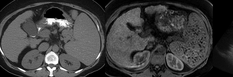
UR Medicine Imaging maintains the newest CT technology, including multi-detector scanners to provide optimum resolution and scanning speed, as well as the latest reconstruction software for multiplanar rendering and volumetric analysis. Examinations range from standard chest, abdomen, and pelvic studies to more complex CT angiography protocols and innovative applications such as cardiac calcium scoring. Our faculty also participate in the interpretation of PET-CT examinations in conjunction with Nuclear Medicine.
Magnetic Resonance Imaging (MRI)
UR Medicine Imaging operates equipment with the latest advances in magnetic resonance imaging technology at our inpatient and outpatient facilities. Advanced pulse sequences, functional MR, MR angiography, and spectroscopy are routinely performed to provide the most accurate diagnostic patient care. We also have an extensive MR research program with an emphasis in cardiac MR and photodynamic therapies.
Ultrasound
UR Medicine Imaging has six sonography rooms equipped with real-time, color flow Doppler, and other state-of-the-art equipment. An additional room is located in the Emergency Radiology annex. The Ultrasound subdivision performs vascular, body, neurosonography, and ultrasound-guided interventional procedures. Although the department has excellent full-time sonographers, residents receive training in the technical aspects of performing ultrasound examinations as a component of a comprehensive education. Faculty and fellows are engaged in clinical and basic science research.
Gastrointestinal & Genitourinary Imaging
The Gastrointestinal/Genitourinary Imaging subdivision provides standard examinations for contrast evaluation of the gastrointestinal and genitourinary tracts. Routine studies include single and double contrast examinations, enteroclysis, three phase pharyngograms for swallowing disorders, excretory urography, cystography, voiding cystourethography and retrograde urethography. Hysterosalpingography is also performed. Faculty and residents also provide interpretation of abdominal and pelvic digital radiographic studies.
Our Faculty