Musculoskeletal
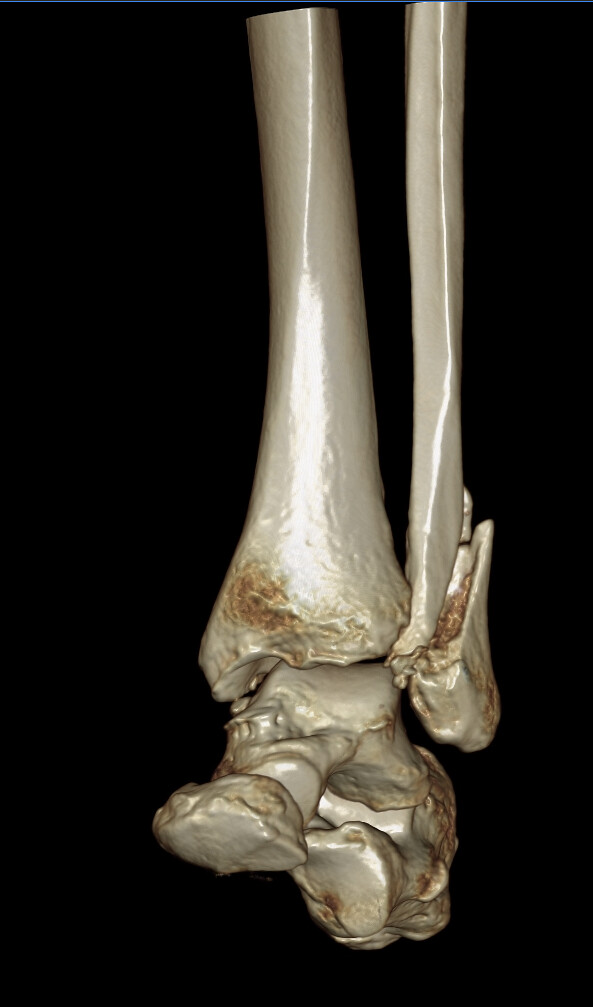
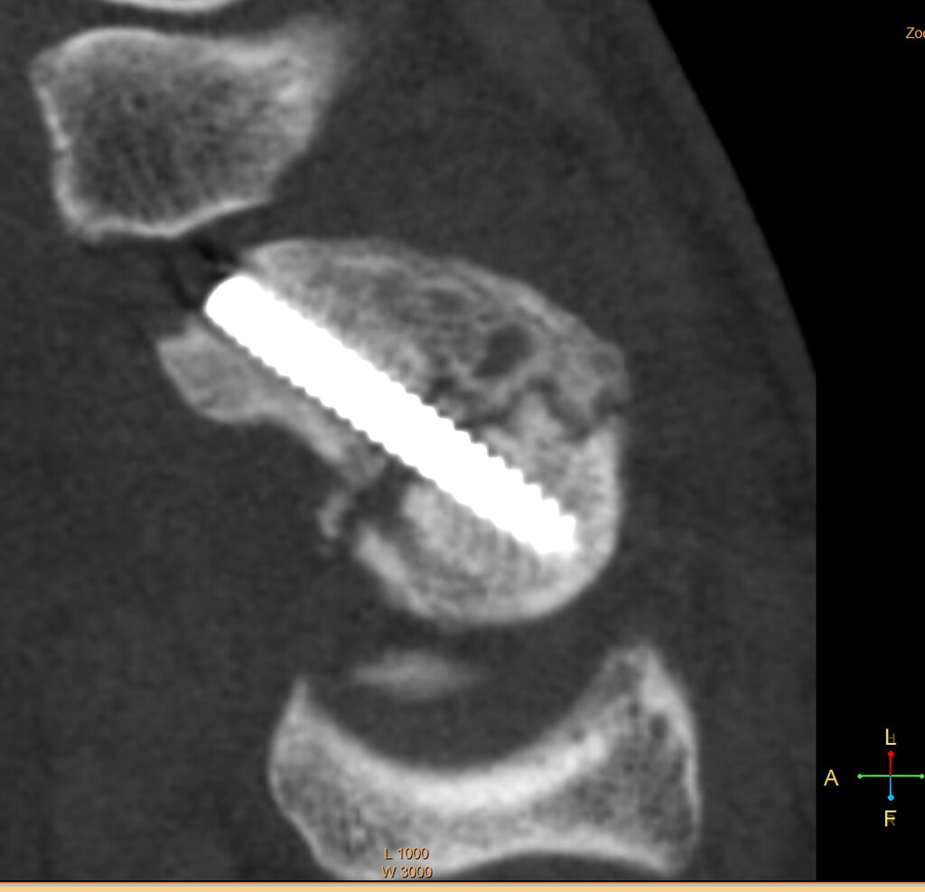
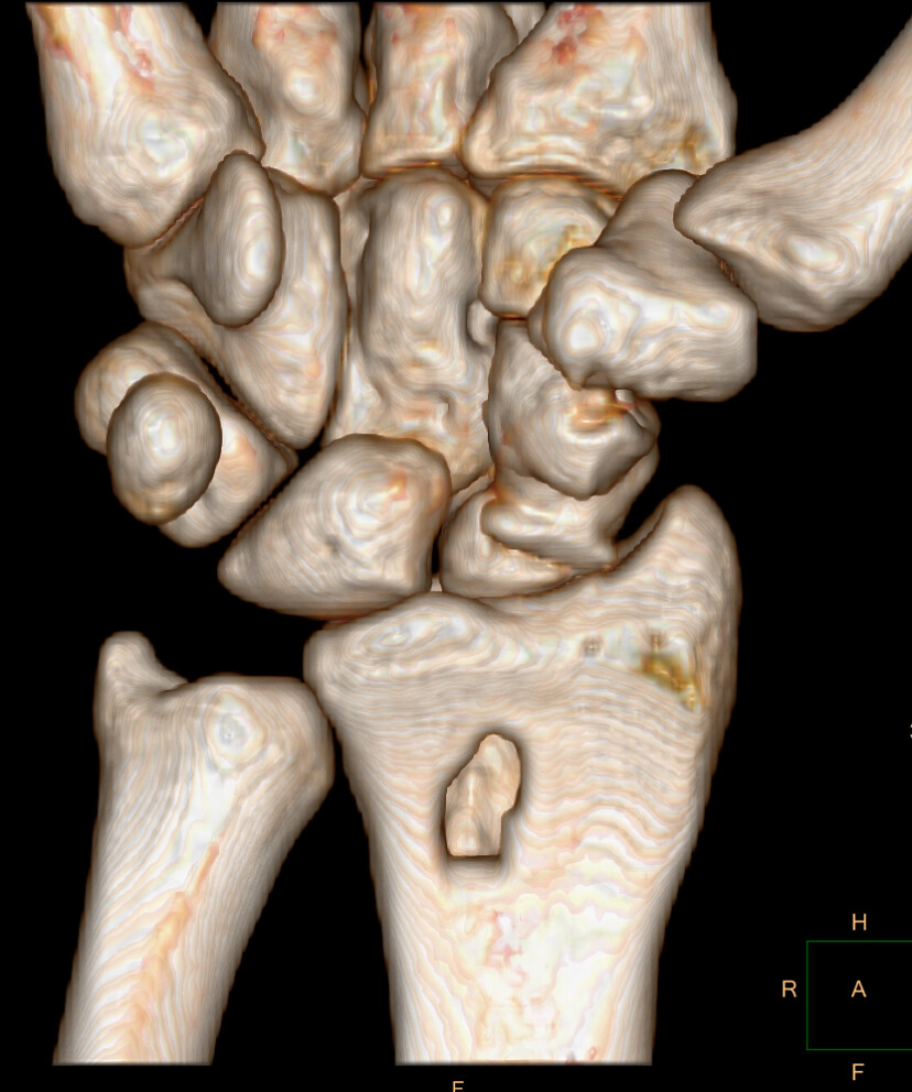
We offer VR and MPR for the:
- Shoulder
- Elbow
- Wrist
- Hand
- Pelvis
- Hip
- Femur
- Knee
- Ankle
- Foot (hind foot)
"The Imaging Sciences 3D lab plays an important role for musculoskeletal CT image reconstruction. In conjunction with the Cone Beam Extremity CT scanner the 3D lab assists in creating consistent and timely 2D and 3D reconstructions with unparalleled image quality when compared to the region. The lab allows for the selection of specialized 2D imaging planes as well as realistic 3D reconstructions which help our orthopedic surgeons when it comes to preoperative planning. The 3D lab also helps create unique imaging reconstructions such as a virtual rotational 3D radiograph which provide valuable information to radiologists and orthopedic surgeons who are in training. Most importantly the 3D lab members take pride in their work and demonstrate initiative in finding ways to continue improving image quality."
- Scott Schiffman, M.D., and Gregory Dieudonne, M.D.


