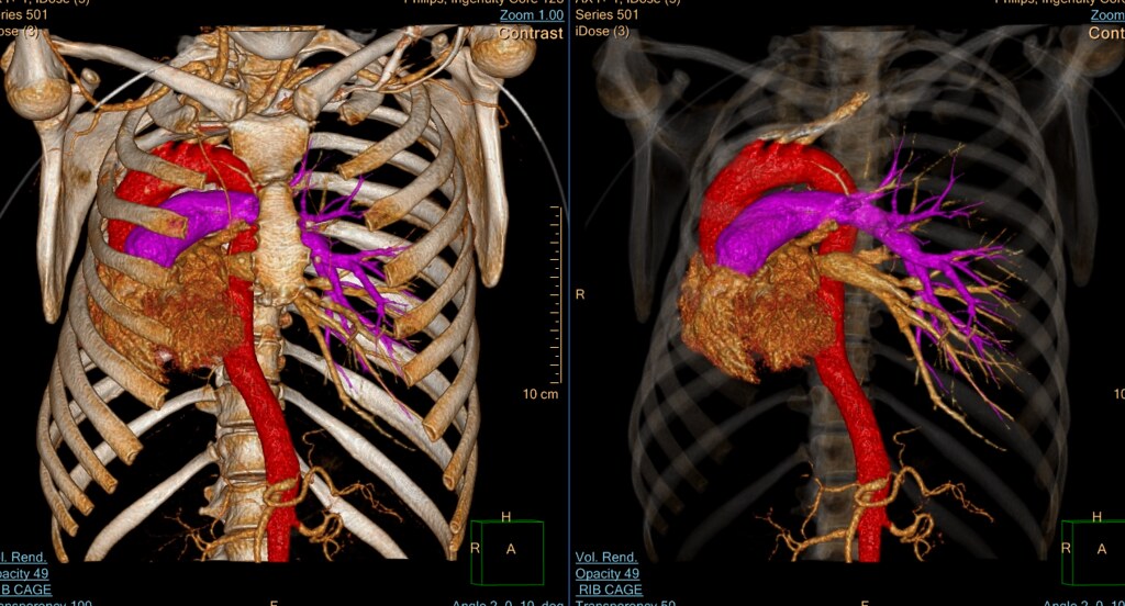
Cardio-thoracic
Cardio-thoracic
We offer CTA of the chest for:
- Pulmonary edema
- Dissection
- Thoracic endovascular aortic repair (TEVAR)
- Left ventricular assist device (LVAD)
- Aneurysms
- SVC clots
- Pulmonary vein mapping
- Transcatheter aortic valve implantation (TAVI)
- Coronary artery stenosis
- Congenital heart
"The 3D lab plays as an integral part in the work of cardiothoracic imaging team. The lab assists us in creating interactive 3-dimensional computer models of cardiovascular imaging, both congenital and acquired, important for both image interpretation as well as preprocedure/presurgical planning by the ordering providers. In addition, the 3-D lab provides initial imaging analysis of these complex studies by identifying and contouring regions of interest, such as the ventricles on cardiac MR imaging and blood vessels, such as the aorta and coronary arteries, on cardiac CT imaging, reducing the overall time needed by the attending for interpretation of these studies."
- Dr. Katherine Kaproth-Joslin


